Tooth resorption in cats
Resorbing teeth are teeth that the body is trying to destroy. They are known as resorptive lesions or kitty cavities. We do not know the cause of this painful condition. The tooth is “eaten” away, exposing the nerve-containing pulp cavity. As time progresses, the tooth may be completely resorbed, leaving just gum tissue where a tooth used to reside. Cats often develop this condition in multiple teeth over a long period of time. This is an extremely common condition and may affect at least 50% of cats during their lifetime.
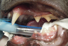
An early sign of this problem is gum tissue starting to move up onto the tooth surface. This can be very subtle and easily missed. More concerning, it is rare for cats to show signs of pain with this condition so owners do not know to have the problem addressed. The condition goes undetected until the pet is brought in for an exam and it is often found when the mouth is examined. When the tooth is probed with a dental explorer under general anesthesia, the jaw will “chatter” due to the exposed nerve in the pulp being contacted.
Often, proof of the pain that was present is seen only AFTER the condition is addressed and the painful teeth extracted. Owners commonly note improved attitude, increased activity levels and more frequent grooming about two weeks after the dental procedure.
Currently, the only treatment for this condition is extraction or crown amputation if the roots of the tooth are completely altered and turning into bone.
Full mouth intra-oral x-rays are essential in treating this condition. It is very common to find multiple teeth affected radiographically that could not be detected clinically even during the oral exam under anesthesia. Cats that have had tooth resorption in the past should be evaluated every year with intra-oral exams and radiographs to detect newly affected teeth before it becomes painful.
Extractions require a surgical approach with drilling of bone over the roots as the affected roots can easily break when extraction is attempted. Cats are given a local dental block and pre-procedure pain medications for any oral surgery performed. All extraction sites are sutured closed with absorbable sutures and cats are placed on pain medication for several days after the procedure. Post-extraction x-rays confirm complete removal of intact roots.
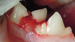
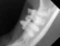
Case #1
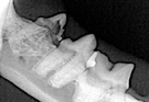
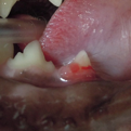
This cat’s lower left Molar has only a mild gingival reaction, but the tooth already has resorption occurring as seen on the x-ray of the tooth on the right. The pulp is exposed so the tooth was extracted.
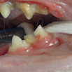
Case #2
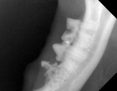
This cat’s premolar (lower jaw, furthest on left) has excess gum tissue over the tooth as the only sign of trouble. On x-ray, it is obvious that the tooth is almost completely abnormal. This tooth was painful and was extracted.